Indian Express
Diagnosed with brain tumour at 38, he returned to cricket, work and normal life
At 46, Amit Mehra* is doing what he loves most — playing cricket, travelling with his family and maintaining a busy professional life as a chartered accountant. Looking at him today, it is difficult to imagine that eight years ago he was diagnosed with a tumour in one of the most critical regions of his brain. “At 38, my whole world had come crashing down. Now I look forward to my weekend cricket match and know when to log out of work,” he says.
Mehra is one of the young Indians reporting brain and Central Nervous System (CNS) tumours in India which have an estimated incidence of 30 cases per 100,000 population. Roughly 30% to 35% of these tumours are malignant (cancerous), while the majority are benign (non-cancerous).
Mehra began noticing brief episodes of weakness and numbness in his left hand while playing cricket. Sometimes he would struggle to hold on to objects. The episodes lasted only a few seconds before disappearing. He attributed them to physical strain, poor posture or fatigue. “It felt like numbness from sleeping awkwardly or overexertion,” he recalls. “The symptoms were so generic that I never imagined they could be related to brain tumour.” Mehra was worried because he kept to a good fitness discipline, ate clean, had no diabetes or hypertension and wasn’t a smoker or alcohol drinker.
That’s the reason he paid attention to his symptoms, which though fleeting in nature, were replicative. An MRI scan revealed a tumour located in the brain’s eloquent cortex — a region responsible for critical functions such as movement and sensation. What appeared to be transient weakness was actually focal motor seizures triggered by the tumour. A surgery and eight years later, there has been no recurrence. His medications have been significantly reduced and he remains active. “I just got a promotion at my firm for high productivity,” he says. His story reflects a larger shift in brain tumour care. Increasingly, a diagnosis that once carried an almost universally grim prognosis is becoming a condition that can often be treated successfully, with many patients returning to work, family life and even sports.
More cases or better detection?
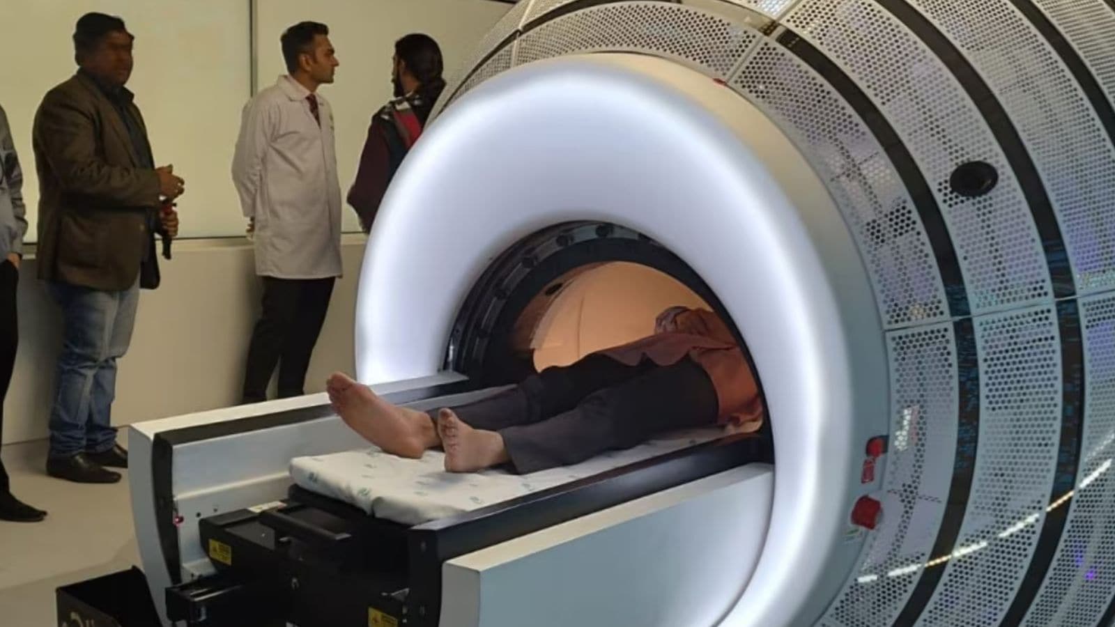
Brain tumours are being diagnosed more frequently in India than ever before. While the incidence remains relatively low compared to other diseases, neurologists say awareness and access to advanced imaging have transformed diagnosis. “Brain tumours are not necessarily increasing dramatically because of new environmental exposure,” says Dr Kapil Singhal, director, Neurology, Medanta, formerly of AIIMS, Delhi. “What has changed significantly is our ability to detect them. MRI scans are more accessible and people are getting evaluated earlier.”
He acknowledges seeing more young and middle-aged adults with brain tumours. Unlike diabetes or heart disease, most brain tumours do not have clearly identifiable lifestyle-related causes. The uncertainty surrounding why they occur often makes the diagnosis unsettling for younger patients who otherwise consider themselves healthy.
Understanding brain tumours
A brain tumour occurs when cells within the brain begin dividing uncontrollably. “It develops when cells in the brain or surrounding tissues acquire genetic mutations that disrupt their normal growth controls. These mutations alter the instructions encoded in DNA, causing cells to multiply and survive longer than they should. As these abnormal cells accumulate, they can form a mass or tumour,” says Dr Singhal.
In most cases, the exact reason behind these genetic changes remains unknown. While some mutations arise spontaneously during a person’s lifetime, others may be inherited from a parent, increasing the risk of developing certain types of brain tumours. “And we need to bust the myth that routine mobile phone usage causes brain tumours. There is no scientific evidence. Also, most brain tumours are not hereditary. They can happen due to random cellular mutations,” explains Dr Singhal.
Contrary to popular belief, not all brain tumours are cancerous. “Many are benign or low-grade lesions that grow slowly and can be completely removed. Modern surgical methods have made longevity and quality of life possible. Low-grade tumours (Grade 1 and Grade 2), which tend to grow slowly have lower recurrence rates. High-grade or malignant tumours (Grade 3 and Grade 4) are more aggressive, have high recurrence chances and require surgery, radiation and chemotherapy,” adds Dr Singhal.
Mehra’s tumour was classified as a Grade 2 lesion — a category associated with relatively favourable outcomes when detected early and removed effectively. “The words ‘brain tumour’ immediately make people think of death or severe disability. But that is simply not true for a significant proportion of patients. Many tumours are treatable, and some can be cured completely,” says Dr Singhal. Apart from primary brain tumours, there are metastatic tumours, too, which spread to the brain from cancers elsewhere in the body.
A revolution in the OR
Perhaps the biggest change over the past decade has been technological. Modern neurosurgery is being driven by sophisticated imaging, computer-assisted planning and real-time monitoring. Before surgery, doctors perform brain mapping using advanced MRI techniques and tractography, which identifies critical nerve pathways responsible for movement, speech and sensation.
“We perform the surgery virtually before doing it real-time,” explains Dr Manish Vaish, director of neurosurgery, Medanta. The imaging data is fed into specialised navigation systems that allow surgeons to plan the safest possible route to the tumour. “The tumour’s location in Mehra’s case made surgery particularly challenging. It was situated in the eloquent cortex, the part of the brain that governs movement and sensation. Even a few millimetres of error could potentially result in permanent neurological impairment,” says Dr Vaish.
Inside the operating theatre, multiple technologies work together. Special dyes can be administered that are absorbed preferentially by tumour tissue, causing the tumour to fluoresce under specialised microscopes. Four-dimensional ultrasound helps distinguish tumour tissue from normal brain tissue in real time. Electrophysiological monitoring continuously assesses the function of critical brain regions during surgery. In selected patients like Mehra, surgeons may even perform an awake craniotomy, allowing communication with the patient during the procedure. “We engaged Mehra by talking about cricket and its statistics. Anything incoherent is a red flag,” recalls Dr Vaish.
He also describes how advanced ultrasonic aspirators have made a delicate surgery procedure easier over the last decade or so. “In simple terms, this surgical tool has a tip that vibrates at very high frequencies (ultrasound), which breaks soft tumour tissue into tiny fragments. At the same time, the device gently suctions away the broken pieces. This is especially useful in brain surgery because tumours are often located close to critical structures that control movement, speech, vision, or memory,” says Dr Vaish. Depending on the complexity of the case, surgeries may last anywhere from three hours to more than 12 hours.
Changing outcomes
One of the most significant advances in neuro-oncology has come not from the operating room but from the laboratory. In the past, doctors relied on what tumours looked like under a microscope. Today, molecular profiling and immunohistochemical (IHC) analysis provide a far deeper understanding of tumour behaviour.
Following surgery, tumour samples are analysed for specific molecular markers and genetic alterations that help doctors predict aggressiveness, recurrence risk and likely response to treatment. “These molecular signatures have become extremely important,” says Dr Vaish. “They help us personalise treatment and provide a much more accurate understanding of prognosis.”
Modern outcomes are no longer dependent on surgery alone. Today’s patients benefit from integrated teams of specialised anaesthetists, critical care specialists, radiation oncologists, rehabilitation experts, nutritionists, psychiatrists and nursing teams.
Back to the mainstream
Recovery timelines vary according to tumour location and complexity. Many patients begin walking within days. Those with low-grade tumours may return to work within weeks. “I went home in three days, worked out of home as I was immune-compromised, walked indoors, reduced screen time to avoid stressing my brain. In three months, I was back at the office and in six months, I was driving again,” says Mehra. Even patients requiring radiation therapy for malignancy resume normal activity during treatment.
Mehra never misses his MRI scans every six months. “That alone has helped me regain something that many patients fear losing after a diagnosis: Normalcy.”
(*Name changed to protect privacy)
View original source — Indian Express ↗
More from Technology

Nepali Telecom
TechnologyJun 8, 2026 · 1 min
Aanchal Kunwar steps down as Daraz Nepal Managing Director
Nepali Telecom

AllAfrica
TechnologyJun 8, 2026 · 1 min
Nigeria: Stakeholders Advocate Local Financing to End Malaria Scourge
AllAfrica

AllAfrica
TechnologyJun 8, 2026 · 1 min
Nigeria: We Are Losing Patience Over Our 42 Abducted Schoolchildren - Askira-UBA Leaders
AllAfrica