
Nature
Lethal plague outbreaks in Lake Baikal hunter-gatherers 5,500 years ago
Main
The analysis of ancient pathogen genomes has significantly expanded our understanding of the evolutionary history of human infectious diseases (for example, Salmonella enterica4 and hepatitis B5), although this has principally been in the context of farming or pastoralist communities. Y. pestis, the aetiological agent of plague, is perhaps the most studied in this regard, and has had devastating consequences on human populations for millennia. Historical outbreaks of plague account for some of the most fatal events in human history1. The recovery of ancient DNA from plague victims has afforded extraordinary insights into the origins and evolution of plague at the time of these events6,7, and, remarkably, revealed infections in prehistoric individuals across Europe8. Historically and today, plague is associated with transmission via fleas from rodents, which successfully adapted to a human commensal niche in the Neolithic9. Genomic analysis of prehistoric plague indicates that in early diverging strains, key genetic adaptations required for flea-mediated transmission of the disease and bubonic infection are absent2,3, leading to uncertainty over the transmission route and severity of these strains.
The detection of early plague cases across multiple generations of Late Neolithic farmers has been used to link outbreaks of the disease to a prolonged demographic decline between about 5,300–4,900 calibrated years before the present (cal bp)10,11, although an alternative explanation attributes the decline to agricultural crisis12,13. The former interpretation has been controversial, with others suggesting infections as more closely resembling benign foodborne enteritis14. The similarity or otherwise of these early strains to Y. pseudotuberculosis—the closest relative of Y. pestis—has been an important point of interest through such discussions, and based on existing ancient genomes, Y. pestis has been estimated to have diverged from Y. pseudotuberculosis some time in the past 50,000 years (refs. 8,11,15).
Studies of prehistoric plague genomes from Late Neolithic and Bronze Age (LNBA) strains predominantly date to between 4700–2400 cal bp (refs. 3,8,16), and are typically defined as one of two lineages, depending on the presence (LNBA+) or absence (LNBA−) of the ymt gene3. ymt encodes Yersinia murine toxin, which enhances bacterial survival in the flea digestive tract during the transition period between rodent and human hosts, and thereby the flea bite-transmitted bubonic form of plague in humans17. Lineages of Y. pestis that diverged prior to these LNBA clades have also been identified in a handful of Neolithic Swedish individuals (5200-4850 cal bp)10,11 and a Latvian individual with western hunter-gatherer ancestry (5300–5050 cal bp)15. These genomes lack classic virulence genes (YpfΦ prophage and ymt), although pangenomic analysis revealed the presence of the locus encoding for Y. pseudotuberculosis-derived mitogen (YPM), a superantigenic toxin associated with Y. pseudotuberculosis (but not later Y. pestis strains). This raises intriguing questions about the possible severity of early strains of plague; subsequent LNBA− strains show substantial gene loss, although the virulence potential of these are unknown3. Evidence regarding the demographic impact of plague infection on prehistoric populations has so far been lacking in these studies.
Middle Holocene hunter-gatherers around Lake Baikal, southeast Siberia, have been the focus of intensive archaeological study by the Baikal Archaeology Project, yielding important datasets for framing prehistoric hunter-gatherer lifeways18,19. These groups demonstrate remarkable continuity of hunter-gatherer lifeways and subsistence, evidenced by an extensive archaeological record of mortuary sites from between about 8500–3500 cal bp (ref. 20). The genomes of sampled hunter-gatherers indicate a long-term continuum of Ancient North Eurasian and North East Asian ancestry until c. 4500–4000 cal bp (refs. 21,22) (Extended Data Figs. 1 and 2). By this period, cases of plague from human remains corresponding to the LNBA− strain are documented sporadically among Early Bronze Age burials22,23. Zoonotic spillover events causing plague infections in this region remain a major health concern to this day24. These are principally associated with marmots, the primary zoonotic reservoir of plague in the region25,26. To explore health and community structure in prehistoric hunter-gatherer groups, we analysed ancient human and pathogen DNA from four cemetery sites in Cis-Baikal (the lake’s western and northern region) across two separate outbreaks dated to 5520–5265 cal bp and 5315–4235 cal bp (95.4% confidence intervals for modelled date ranges based on individuals with detected plague cases, corrected for freshwater reservoir effects; Supplementary Note 4). The long tail for the second outbreak date range (Fig. 1b) is due to this only consisting of two direct dates, although the highest likelihood date range for this is approximately 5050–4850 cal bp.
Outbreaks of basal plague strains
We generated shotgun-sequenced ancient DNA from 46 Late Neolithic individuals and examined this data for presence of pathogens (Methods). This revealed a conspicuously high occurrence of Y. pestis among these individuals, more so than any other pathogen. Y. pestis was detected in 18 individuals, indicating 2 distinct phases of outbreaks of plague infection—separated by between 4 and 6 centuries—in 4 cemeteries (Fig. 1a,b). These occur across two phases at Shumilikha, Ust’-Ida I, Bratskii Kamen and Serovo (see Fig. 1c), with cases from Bratskii Kamen in both the first and second phases. These sites are all located on banks of the River Angara, a major watercourse draining from Lake Baikal, with a rich fishery27. Stable carbon and nitrogen isotopic data from individuals at Ust’-Ida I evidence consumption of both local fish and terrestrial game27. Burials at Ust’-Ida I and Shumilikha correspond to the Isakovo mortuary tradition (characterized by bodies that are typically oriented parallel to the river, and the presence of grave goods such as mitre-shaped clay vessels, lithic arrowheads and bone or antler points), whereas those at Bratskii Kamen and Serovo correspond to the Serovo mortuary tradition (with bodies frequently oriented perpendicular to the river; bifaces and egg-shaped pots as grave goods are the main features; see also Supplementary Note 1). At Ust’-Ida I, we also detect reads aligned to the zoonotic pathogen Brucella, the cause of brucellosis, in one individual (#26.04; Supplementary Note 3). The two plague outbreaks are grouped by the predominant burial practices at each cemetery: Isakovo-style graves in the first outbreak and Serovo-style graves in the second phase (Supplementary Note 1), which are contemporaneous at Lake Baikal between around 6000–5000 cal bp (ref. 27). This period is defined locally as the Late Neolithic, following Siberian archaeological terminology, where the Neolithic is defined on technological criteria such as the introduction of the bow and arrow, clay vessels and stone grinding techniques (domestic plants and animals other than dogs are absent), although these communities remain as hunter-gatherers until the encroachment of pastoralism in the Late Bronze Age. Grave sites comprise the vast majority of the Cis-Baikal archaeological record, and designations such as the Late Neolithic are later categorizations, applied to distinct sets of burial characteristics and grave goods that broadly correspond to different periods. All four cemeteries were also used during the Early Neolithic (7650–6660 cal bp) and Early Bronze Age (4970–3470 cal bp)27, although only their Late Neolithic components are considered here.
Pairwise sharing of identity-by-descent (IBD) segments between individuals at these cemeteries indicates recent shared ancestry. Although they are up to 340 km apart, the Angara river would readily have facilitated travel. Very low rates of inbreeding were detected and a high effective population size based on runs of homozygosity was inferred using hapROH (maximum likelihood estimate: 18,219 individuals, 95% confidence interval 9,445–42,062). This is consistent with the scenario of highly mobile, exogamous hunter-gatherer groups.
Within the hunter-gatherer individuals analysed here, the highest number of detected plague infections was at Ust’-Ida I, which is also the largest Isakovo mortuary site in Cis-Baikal. Here, we found a 35% detection rate (11 out of 31 individuals sequenced), including burials #14 and #56.01, for which human genome data were previously reported21. Across other sites, we identify one high-coverage plague genome at Shumilikha, four lower coverage genomes from Bratskii Kamen, and one medium coverage genome from Serovo. Overall, we observe a 39% detection rate across Late Neolithic individuals at these cemeteries (from dental cementum). In comparison, quantitative PCR screening of known Mediaeval plague victims at Smithfield, London, UK28 returned a detection rate of 5.7% from bone and 37% from dental pulp tissue (overall 20%), indicating a high rate of false negative plague detection using ancient DNA. To prevent misrepresentation of data, all ancient individuals with screening data from the affected sites are reported here (human autosomal genome coverage ranges from 0.001× to 1.9×, average 0.65×). Direct radiocarbon dates were obtained from nearly all the individuals within the Late Neolithic components of these cemeteries (a total of 58, including those previously reported from Ust’-Ida I29; Supplementary Data 7).
Y. pestis genomes identified between the two Baikal phases of outbreaks were found to diverge ancestrally to the current known clade of ancient and modern plague strains (Fig. 2). We confidently assign these to Y. pestis from their phylogenetic position, and also the presence of virulence genes and plasmids characteristic of Y. pestis (Extended Data Fig. 3 and Supplementary Note 3). This phylogeny was built using genomes obtained from Shumilikha Burial #34 (6.4× coverage) from the first phase, and from Bratskii Kamen Burial #22 (1.6×) and Serovo Burial #10 (1.0×) from the second phase. Eight lower coverage genomes were phylogenetically placed using UShER30. The UShER algorithm finds the most parsimonious placement on the tree, selecting the node with the greatest number of descendents if multiple are equally parsimonious, and ignores missing genotypes. Placement of all low-coverage genomes at the same basal node is partly due to data missingness, although consistent with the position of the three higher coverage Baikal genomes. Bayesian inference of node dates was undertaken following an approach to account for the effects of recombination within bacterial phylogenies31,32 (Methods and Supplementary Note 3). The emergence of Y. pestis as a clonal species of Y. pseudotuberculosis occurs some time between the divergence of the lineage that gives rise to Y. pestis (labelled node A in Fig. 2) and the most recent common ancestor of available Y. pestis genomes (node B in Fig. 2). The upper bound provided by the former is likely to be substantially affected by the paucity Y. pseudotuberculosis genomes sequences, and could well be more recent if phylogenetically closer serovars were identified. Nonetheless, this lower bound (with a mean date of 5,709 years ago) revises a previous divergence estimate of 4,810–5,122 years ago33, as would be expected by including Y. pestis genomes older than this range (other estimates of this have ranged from 6,000 to 50,000 years ago8 and 7,400 years ago15). The phylogeny supports the conclusion that Y. pestis first evolved from a variant of the O:1 Y. pseudotuberculosis strain (represented by a genome from serotype O:1c, European Nucleotide Archive (ENA) accession: SAMEA7160327), consistent with previous findings34 reporting the inactivation of the O-antigen gene cluster as a step towards the evolution of Y. pestis. Between the two phases, we observe small genetic differences between strains in distinct private mutations in the first and second phase strains (with strict filters for genotype calling; Methods and Supplementary Note 3); this is also clear from the position of nodes in Fig. 2. Although mutation rates in Y. pestis are known to be highly variable within different lineages33, this result is consistent with a scenario of related strains resulting from separate zoonotic spillover events from a local animal reservoir.
Baikal hunter-gatherer plague mortality
To contextualize these plague outbreaks, we considered biological kinship patterns, burial treatment and age at death within the affected hunter-gatherer cemeteries. At the site with the highest positive detection of plague (and largest sample), Ust’-Ida I, radiocarbon dates for the Late Neolithic Isakovo component are exceptionally tightly clustered for a relatively large cemetery29 (Extended Data Fig. 4). Modelled date ranges for all the early phase plague victims indicate a very narrow temporal span, on the order of a few decades (Supplementary Note 4), supporting the scenario that these burials were contemporaneous. This is further corroborated by the high similarity among plague genomes consistent with plague infections occurring in a single outbreak, or over a very brief time span. By reconstructing the most likely familial pedigrees, we find that the relationships and ages of family members are consistent with a mortality event over a time span of less than a single generation (Fig. 3). None of the age at death–relationship pairings indicate, for example, children that reached a similar age to their parents, or siblings and half-siblings with very different ages (the greatest sibling age gap is nine years, separated by a middle sibling). Where multiple generations are present, their inferred age-at-death ranges are generally consistent with those expected if all relatives had died at the same time (for example, a 12–15 year old has a 35–50-year-old father).
The Isakovo mortuary group at Ust’-Ida I is unusual in several other ways among Cis-Baikal hunter-gatherer cemeteries. In addition to the tightly clustered radiocarbon dates, childhood mortality is disproportionately high (also observed at Bratskii Kamen, see Fig. 4), and there is a high incidence of multiple-interment graves (more than half at the site) with no evidence of subsequent grave opening and addition of new burials. This suggests co-occurrence of deaths within shared graves, consistent with a catastrophic mortality event. An avuncular relationship was also detected using KIN35 between the Ust’-Ida I and Shumilkha cemeteries, but this was not substantiated by the expected IBD-sharing pattern (Supplementary Note 2). Nonetheless, the high degree of IBD sharing between individuals (Fig. 1) across a distance of only 37 km along the Angara river, suggests that the concurrent plague outbreaks might be linked to the groups being in close contact at this time point.
In terms of plague detection within grave groups, we find no statistically significant pattern of plague co-occurrence among relatives (Supplementary Note 3), although affected individuals appear to be associated in a number of cases. The burial at Bratskii Kamen features a shared grave of 3 young girls, aged between 4 and 9 years (Fig. 3, left), with similar radiocarbon dates (Supplementary Note 4). Two of them (#19.01 and #19.03) were inferred as third-degree related (most probably cousins); the third had insufficient DNA preservation to confidently infer relatedness, but all three shared a mitochondrial haplotype with three rare private mutations and so were likely to be close maternal relatives. Genome data for Y. pestis were identified in all three, suggesting an outbreak of plague infection in a family, with synchronous deaths of the three children. Similarly, at Ust’-Ida I, a nephew and aunt (#20.01 and #20.02) are buried in a shared grave, with Y. pestis identified from both (Fig. 3, orange pedigree). The teenage niece of the aunt, however, is buried in a different shared grave with an unrelated teenage male (possibly suggesting non-biological kinship); his father in turn (green pedigree) is buried in an entirely separate grave.
Additionally, some pairs of siblings who are buried together in shared graves show only one individual detected as positive for plague, as is the case with the siblings in grave #25 (Fig. 3, red pedigree). In another example, for a sister (#26.01) and brother (#26.04), the sister is inferred as positive, whereas the brother is not (although Y. pestis reads are detected at just below the threshold for confident identification; Supplementary Data). These observations are consistent with a high false negative detection rate in the palaeogenomic analysis of plague28. The brother was also infected with probably non-lethal brucellosis (Supplementary Note 3). In several cases close family members are found in different graves within the cemetery, for example, Burial #8, the third sibling of the pair in Grave 26. A pattern is visible, where two closely related family members are buried together, and one or more others are buried further away. This may be consistent with a more drawn-out sequence of deaths instead of a single mortality event if shared graves indicate concurrent deaths, reflecting a scenario of delayed person-to-person disease transmission. No causes of death were apparent other than genetically detected plague infection (although other microbes detected might reflect bacterial coinfections at the time of death; Supplementary Note 3). Notably, survivors must have existed to bury the deceased, with the typical Isakovo mortuary treatment and grave goods as well as acknowledgement of biological kinship suggesting a more prolonged sequence of mortality events.
Epidemiological implications
At Baikal, the principal contemporary zoonotic reservoir of plague is the marmot (Marmota sibirica), and marmot hunting for meat and fur has historically resulted in perennial plague infections especially in young men, who are exposed during skinning and butchery36. Since the nineteenth century, marmots were the most targeted game species by Indigenous hunters in this region, originally by trapping37, and there are extensive historical accounts of ‘tarbagan plague’ from consumption of infected marmots around Lake Baikal38. Prehistoric hunter-gatherer marmot procurement is clearly evidenced by the presence of numerous marmot teeth as grave goods in Early Neolithic Kitoi graves19,39, although these have not been found in Late Neolithic graves. Consumption of raw or undercooked marmot organs results in the septicaemic form of infection following the faecal–oral transmission route, whereas close contact with marmots infected by present-day Y. pestis strains causes bubonic or pneumonic plague (or often both), with the latter often occurring secondarily to septicaemic infection40 or inhalation of infectious blood droplets during, for example, skinning41. The incidence of detected infections among co-buried kin described above would be consistent with the transmission of plague among humans, particularly via pneumonic transmission in the scenario of concurrent deaths.
A striking aspect of the osteological age-at-death data at Ust’-Ida I and Bratskii Kamen—the two cemeteries with multiple instances of plague detected—is that their demographic profiles are highly skewed towards childhood mortality. These both show a peak in mortality at the age range of 7.5–11 years—that is, in children before puberty (Supplementary Note 1). In an analysis of mortality profiles across mid-Holocene Cis-Baikal hunter-gatherer cemeteries, these two cemeteries are clearly outliers in terms of the proportion of childhood deaths (Fig. 4). This result was found to be highly statistically significant given a null model of mortality profiles (Supplementary Note 5). Conversely, the 20–25-year age range shows the lowest mortality at Ust’-Ida I, and deaths between 20–35 years of age are completely absent at Bratskii Kamen (Supplementary Fig. 4). Parents are also conspicuously absent from the pedigree groups; although there are many sibling and cousin relationships, there is only one instance of a parent–offspring relationship. The sex ratio in these individuals appears unaffected however (22 XY and 24 XX).
In the context of widespread infection with a plague strain of unknown virulence, this differential mortality between children and adults might be interpreted in a number of ways, given the available bioarchaeological data and current understanding of human immunity. First, adults could largely consist of those who had already been exposed to and recovered from the plague as children, and consequently acquired protective immunity, preventing reinfection or death. This would imply that outbreaks were regularly recurring, which our findings are not able to attest to, and would also imply that older individuals would be more likely to have acquired immunity, yet mortality actually increases slightly after the age range of 20–35 years (after the primary peak around 10 years of age). Alternatively, variation in mortality due to behavioural differences between age groups (for example, division of group tasks or roles by age, resulting in higher childhood exposure to marmots) cannot be ruled out, although there is little analogous precedent for this with regard to marmots specifically, and this is not supported by the lack of heightened childhood mortality in any other Baikal hunter-gatherer cemeteries (Fig. 4). Finally, it is possible that children could be at a greater risk of death owing to inherent differences in immune responses between adults and prepubescent children. Children are known to be more susceptible to infection from Gram-negative bacteria42, as evidenced by the epidemiological profile of Yersinia enterocolitica and Y. pseudotuberculosis infections today43.
Functional variants in basal Y. pestis
The evolution of Y. pestis lineages is shaped considerably by processes of gene loss44, a pattern typical of pathogenic bacteria in the transitional process to obligate parasitism, which has also been identified across the LNBA− plague strains3. From analysis of the coverage of the classic plague virulence genes, we find that virulence genes absent in published LNBA− and pre-LNBA strains from Riņņukalns (RV 2039) and Falbygden11 are also absent at Baikal (ymt and YpfΦ prophage; Fig. 5), prohibiting the manifestation of bubonic plague (Extended Data Fig. 3). However, since this virulence gene analysis relies on traditional single-reference mapping, it is restricted to genetic content that is present in the modern reference. To characterize possible ancestral Y. pseudotuberculosis variation in the Cis-Baikal strains which might contribute to our interpretation of their pathogenicity, we mapped sequenced reads to a pan-genome variation graph representing genetic diversity across 82 complete assemblies of the Y. pseudotuberculosis species complex (56 Y. pestis, 24 Y. pseudotuberculosis and 1 Yersinia similis, based on ref. 11). We found that the two plague strains from Lake Baikal carried similar levels of ancestral Yersinia diversity that were only found in Y. pseudotuberculosis and Y. similis as other pre-LNBA strains (Fig. 5e). For example, we detected the presence of ypm, the gene encoding the YPM superantigen known from modern-day Y. pseudotuberculosis strains45, and recently observed in pre-LNBA and LNBA− plague strains11. Three alleles of this gene exist in modern Y. pseudotuberculosis: ypmA, ypmB and ypmC, with ypmA being regarded as the most virulent form of the gene46.
YPM binds to the invariant region of human leukocyte antigen (HLA) class II molecules and interacts with the variable domain of the β chain of the T cell receptor. By bridging HLA class II and a T cell receptor, YPM promotes T cell activation and the release of a range of proinflammatory cytokines, further amplifying the immune response47,48.
These YPM-associated immune responses have been suggested to be the cause of various inflammatory complications, including encephalopathy, Far East scarlet-like fever (FESLF; also known as Izumi fever; especially associated with ypmA) and a Kawasaki-like syndrome47,49,50,51. Today, FESLF occurs primarily in children aged less than 14 years and Kawasaki disease occurs mostly in children aged 5 years or less. However, Kawasaki disease following Y. pseudotuberculosis infection may also affect older children52. These YPM-related inflammatory complications are likely to also have primarily affected prepubescent children in the past, further exacerbating the early Y. pestis induced morbidity and mortality in the young.
Notably, we find that the ypm gene from the two plague strains from Cis-Baikal is closest in sequence similarity to ypmA, differing at only 3 base positions: 4653 (T>G (isoleucine>arginine)), 4711 (C>T (synonymous threonine)) and 4770 (G>A (glycine>glutamate); Fig. 5a). These three single nucleotide polymorphisms (SNPs) appear to be fixed in all plague strains in which the gene is present (pre-LNBA and LNBA− strains; Fig. 5a). Since two of these three variants are non-synonymous mutations (I54R and G93E are located in distinct beta-sheets of the YPM structure), they could potentially influence protein secondary, tertiary or quaternary structure, protein–protein interactions, and recognition by the immune system (Fig. 5b). Furthermore, by reconstructing the most likely phylogeny of the three known ypm variants together with our data, we found that the ypmB version of the gene is highly divergent from ypmA, ypmC and the ancient plague ypm. Under the assumption that the root of the tree is located between ypmB and the remaining diversity, the ypm from ancient plague appears to diverge ancestrally to both ypmA and ypmC (Fig. 5c).
In addition, we identified ten open reading frames (ORFs) around the ypm locus that are present in the ancestral form of the plague but are absent in later forms. The ORF region is similar to an unstable region of the Y. pseudotuberculosis genome with notable low GC content. We found that these ORFs in the Baikal Y. pestis genomes were similar to those surrounding the ypmB variant as reported in other pre-LNBA strains11 (Fig. 5d). This pattern with a ypm gene similar to ypmA combined with a ypm locus similar to ypmB has—to our knowledge—not been observed previously. A possible explanation could be that this diversity in the Lake Baikal pre-LNBA strains, the most basal of the sampled plague strains, reflects ongoing local adaptation to marmots and other rodent hosts to a greater extent than humans because the regional animal host reservoir presumably at this time far exceeded that of humans. This, so far unique, combination might affect, for example, gene methylation and ypm transcription levels.
These genetic features of the Baikal pre-LNBA strains might, together with age-dependent differences in the immune system, partly help explain why prepubescent children predominate among the plague victims, although assessing their actual impact requires functional studies.
Discussion
Our findings demonstrate that the earliest known outbreaks of plague occurred in prehistoric hunter-gatherers centuries before infections are observed in Neolithic farmers. These outbreaks were probably the result of zoonotic spillover from wild marmot populations at Lake Baikal. These results support a central or northeast Asian origin for plague, whereas previously the earliest samples had only been reported in northern Europe11,15. This is in line with estimates based on the analysis of modern Y. pestis diversity53. Our phylogenetic analysis reveals that these virulent plague strains are temporally relatively close to the most recent common ancestor of Y. pestis and Y. pseudotuberculosis, possibly indicating rapid diversification with the transfer to rodent hosts from one or several of the other animal hosts of Y. pseudotuberculosis. Additionally, this raises questions around the differentiation of taxa within the Y. pseudotuberculosis species complex (which includes Y. pestis and Y. similis) that ancient genome data alone may not be adequate to answer (given that conventional distinctions can be based on pathogenic potential and host range as well). Furthermore, the inferred high lethality of these outbreaks is directly evidenced by mortality profiles and coinciding radiocarbon dates in affected burial sites, indicating that children and adolescents were especially vulnerable; these are insights that were previously lacking for prehistoric plague infections. Until now, the earliest detected strains of plague were of uncertain pathogenicity; their virulence has been the focus of considerable debate, based on genetic data alone3,10,15,16,54. Here we have integrated multiple lines of evidence from mortuary sites affected by plague (including plague genomes, biological kinship patterns, mortality profiles and modelled radiocarbon date ranges) to characterize what we argue are the lethal consequences of infection during this period.
The context of these outbreaks is important for the interpretation of health and epidemiology in the past. That these outbreaks occur in relatively small, mobile prehistoric hunter-gatherer groups emphasizes that increased population density, animal domestication and lifestyle changes resulting from the Neolithic transition are not necessary conditions for significant zoonotic outbreaks. This further revises interpretations of plague as a unique factor contributing to demographic decline during the Late Neolithic in Europe, as previously suggested10,11, especially given the apparent severity of outbreaks identified here. The mortality profile at Baikal also contrasts sharply with the expectation of proponents of the Neolithic epidemiological transition theory that the greatest burden of zoonotic disease in prehistoric hunter-gatherers would fall on producers (20 to 40 year olds)55. Our findings further reveal insights into the social dimension of these communities during outbreaks, evidencing care for the dead (from the co-interment of close relatives and apparently contemporaneous victims), and concurrently detected infections among kin which evince interpersonal contact in life. This evidence of person-to-person transmission contrasts with previous expectations for basal plague strains15.
Of note, children seem to have borne the brunt of lethality from plague infections at Cis-Baikal. Different mortality rates among age groups have been observed in historical records of plague outbreaks. Similar to our findings, Parish records from the bubonic plague outbreak in London (UK) in 1603 showed a considerably higher child mortality rate (around 5× higher)56,57. One distinct difference between the Cis-Baikal outbreaks and mediaeval bubonic plague epidemic is the probable transmission route. Although airborne and faecal–oral transmission might have occurred in both, flea bite transmission associated with bubonic plague is unlikely in the Cis-Baikal outbreaks (given the absence of ymt). Spread of infectious droplets or aerosols through coughing is documented as the primary transmission mode of pneumonic plague58, matching our findings for human-to-human spread inferred from the biological kinship and archaeological data. Notably, our results are consistent with previous interpretations that early Y. pestis strains could have presented as fatal respiratory pathogens54.
Our results indicate that the earliest observed zoonotic spillover was not a one-off event but re-occurred several centuries later, highlighting the prominence of zoonotic infections in prehistoric communities across many different cultural and environmental settings. Additionally, a low-coverage identification of brucellosis suggests evidence of animal-to-human zoonotic transmission in these groups (infection is acquired by direct contact with infected animals59, Supplementary Note 3). Recurrent outbreaks of ancestrally diverged plague strains in Cis-Baikal groups between 5500–5000 cal bp further suggests a long history of wild rodents as a perennial reservoir for plague spillover. The subsequent plague strains genetically closest to those at Cis-Baikal are from around 5,000 km west in northern Europe. Given this distance, and that there is little evidence for external contact with non-hunter-gatherer groups at this time, this supports the hypothesis that a substantial, continent-spanning rodent reservoir of Y. pestis might account for frequent isolated spillover events in the subsequent millennia, instead of continuous human-to-human transmission. Moreover, the potential association of prehistoric spillover with human procurement of marmots at Cis-Baikal emphasizes the probable key role of rodent species other in the composition of Y. pestis reservoirs. Some 352 reservoir species have been identified from present-day surveillance, a number of which are ecologically long established (for example, ground squirrels and gerbils)60. The scenario of a persistent prehistoric reservoir aligns both with previous findings of rapid repeated infections of diverged plague strains within the same familial lineage from Neolithic Sweden11 as well as at Cis-Baikal. We note that recent findings show that early plague cases in domesticates61,62 may equally be congruous with reverse zoonosis, although given the long history of rodent species reported as reservoirs for plague60, we feel that this currently remains the most parsimonious source for these outbreaks.
Together, our findings underscore the universality of zoonotic infection, given the markedly different lifeways of prehistoric hunter-gatherers from European Neolithic farmers. These insights are as relevant for the challenges faced by the world today as they were 5,500 years ago, with 75% of new human pathogens emerging from animal transmission63. Insights into the evolutionary history of pathogens across periods of substantial demographic and technological change (before the impact of the Neolithic transition in this case) can provide data to contextualize the major challenges humanity is currently facing, such as the climate change-driven disruption of ecological niches around the world64.
Methods
Laboratory work
Ancient DNA was extracted from the dental cementum of molar or premolar teeth from archaeological skeletal remains studied by the Baikal Archaeology Project. Sampling for ancient DNA (aDNA) was undertaken in dedicated clean laboratory facilities at the Lundbeck Foundation Centre for GeoGenetics (Copenhagen) and at the Institute of Archaeology, University College London (London). Cementum was isolated specifically from the roots of teeth67 using a sterilized handheld rotary saw, and pulverized prior to demineralization and enzymatic digestion. Sampled aliquots were approximately 50–100 mg of material. Extraction, purification and library preparation of aDNA for shotgun sequencing followed the approach described in Allentoft et al.68, using a double-stranded library protocol following Margaryan et al.69 in the first instance, and the ‘Santa Cruz Reaction’ single-stranded library protocol70 for samples with low template DNA content. Concentrations for resulting libraries were obtained using an Agilent FragmentAnalyzer and pooled at equimolar concentration for sequencing on Illumina NovaSeq 6000 S4 flowcells (100 bp paired-end reads) at the GeoGenetics Sequencing Core (Copenhagen). All samples were screened without partial Uracil-DNA Glycosylase (UDG) treatment, and in some cases subsequent additional libraries were built with UDG treatment (following71).
For libraries where screening sequencing indicated the presence of Y. pestis DNA, in-solution capture enrichment was undertaken. Hybridization capture was performed using the Arbor Sciences myBaits kit following Wagner et al.72, using the manufacturer’s High Sensitivity protocol, but only with a single round of enrichment. Pooled libraries from the capture reactions were then re-amplified for 16 cycles and sequenced on the same platform as above.
Preliminary bioinformatics
Following base-calling of Illumina data using CASAVA (v.1.8.2)73, adapter sequences and polyN tails were trimmed from demultiplexed fastq files using AdapterRemoval (v.2.0). Reads were aligned to the human reference genome GRCh38 using bwa aln (v.0.7.18)74 (reference genome hg19 was also used for hapRoH analysis, see below). Aligned reads were converted to BAM files, merged across libraries at sample level, sorted, filtered and indexed using Samtools (v.1.21)75, then duplicates identified using MarkDuplicates from Picard (v2.18.7), with the following options in place: ‘OPTICAL_DUPLICATE_PIXEL_DISTANCE = 12000 REMOVE_DUPLICATES = false TAGGING_POLICY = All VALIDATION_STRINGENCY = LENIENT’. Duplicate reads were then filtered out using Samtools alongside reads with a mapping quality of <30. Summary statistics for sequencing depth and coverage were generated using BEDtools (v2.23.0)76 and pysam (https://github.com/pysam-developers/pysam). Estimation of human DNA contamination and damage patterns were performed at a library level, using contamMix77, ANGSD (v.0.940)78, and mapDamage2.079.
Human DNA Analysis
Chromosomal sex was inferred based on the ratio of Y and X chromosome aligned reads, following existing confidence intervals80. Chromosomal aneuploidies were not detected. Mitochondrial haplogroups were assigned using haplogrep (v.2.4.0)81 following annotation of variants using mutserve (v.1.3.0)82. Y chromosomal haplogroups were assigned following the approach in ref. 11.
For exploratory analysis of ancestry through principal component analysis (PCA), pseudohaploid genotypes were called by randomly selecting a variant from a pileup generated with Samtools. Samples were then projected into the variation space obtained from using smartpca83 to undertake PCA on 2,086,279 SNPs (filtered for transversions only and with minor allele frequency >0.1%) from a reference panel of ancient Eurasian populations68. The latter was lifted over from hg19 using hgLiftOver (https://genome.ucsc.edu/cgi-bin/hgLiftOver), and the effects of liftover evaluated (Extended Data Fig. 2). All reported samples were included in the PCA by projection.
Diploid genotypes were called using bcftools (v.1.21), and for the analysis of IBD segment sharing, missing diploid genotypes were imputed using GLIMPSE84 (for samples with a minimum autosomal genome coverage of 0.1×) following the approach in ref. 68, and IBD segments called using IBDseq (v.r1206)85, followed by genetic clustering by IBD68.
Runs of homozygosity were detected from homozygous-by-descent segments obtained from IBDseq, and from pseudohaploid data subset to the 1240k SNP positions using hapRoH (v.1)86. Biological kinship was inferred using KIN35, initially running KINgaroo on filtered BAM files targeting the 2,086,279 SNPs described above, and then validated based on IBD sharing inferred from IBDseq. Pedigrees were then reconstructed taking into account the resulting log-likelihood estimates for kinship scenarios, uniparental haplotypes, sex and age-at-death (Supplementary Note 2).
Screening for pathogen taxa
Shotgun sequencing data generated from dental cementum was screened for the presence of known human pathogens using the pathopipe workflow (https://github.com/martinsikora/pathopipe/) detailed in Sikora et al.87. Reads were classified using a fast k-mer approach, KrakenUniq88 (v.0.5.8), based on a custom database of human pathogens and environmental microbes. For each genus identified in each sample, pairwise alignments using bowtie289 (v.2.5.4) were made for all reads classified to that genus against all available species reference genomes for the same genus. Assignments are then made for the presence of pathogen taxa on the basis of the following detection thresholds: unique read count >30, k-mer rank = 1, corrected coverage ratio >0.5, average nucleotide identity >0.97, average number of soft clipped bases <8, based on those applied by Seersholm et al.11.
Y. pestis DNA analysis
For samples where plague infection was identified through the pathogen screening pipeline, we carried out traditional single-reference mapping with Bowtie289 against the plague reference genome (CO92; GCA_000009065.1), with the parameters ‘-D 20 -R 3 -N 1 -L 20 -i S,1,0.50–end-to-end–no-unal’. Next, duplicate reads and low mapping quality reads (MQ < 30) were removed with samtools75, followed by calculations of the average depth of coverage in each sample using BEDtools genomecov76. We characterized plague cases based on their coverage as either: tentative detections (<0.01×), lower-coverage plague cases (0.01-1×) or higher-coverage plague genomes (>1×).
For the three higher coverage genomes we called genotypes in a sample-wise manner using HaplotypeCaller from GATK90, followed by a subsequent step of joint haplotype calling using GenotypeGVCFs on the merged dataset. Using VariantFiltration (GATK) we removed low-confidence calls of either: low genotype quality (<50), an allele balance of less than 0.9, a read depth of less than 3 or a read depth higher than 1,000. Next, we converted the dataset to multifasta format using bcftools consensus. In doing so, we applied a mask across regions containing the highest proportions of zero mapping quality reads, typically localized in repetitive regions, following the approach in Seersholm et al.11. To maintain the coordinates of the reference genome and for consistency with the other aligned samples, sequencing data for published reference genomes for Y. pseudotuberculosis and Y. similis were downloaded from ENA and the reads were realigned to the Y. pestis reference genome GCA_000009065. Genotypes were then called and filtered as described above, yielding a multiple sequence alignment for the full Yersinia chromosome with 448 sequences on the coordinates of the reference genome. A phylogenetic tree was inferred from the full alignment file including all reference sequences and the three high-coverage samples from this study using RAxML-NG91 with the GTR + G substitution model and using the Y. similis reference genome (SAMEA5779183) as an outgroup (see Extended Data Fig. 3). We converted the multiple sequence alignment to a haploid VCF using faToVcf30 with the Y. pestis reference genome NC_003143.1.fa as a reference, then built a mutation-annotated tree object from this VCF and the RAxML phylogeny using UShER30. This new UShER phylogeny maintains the original topology but directly assigns substitutions in the VCF to branches on the tree using the Fitch-Sankoff algorithm92,93, so that edge lengths are in units of real substitutions. We used matUtils94 to extract a.json file from the mutation-annotated tree protobuf, available interactively online at https://bit.ly/Ypestis_MAT.
For the 8 lower coverage samples, we called a SNP-only vcf using bcftools, filtering for a minimum mapping quality of 30 and bases, a minimum base quality of 30, and a maximum depth of 1,000. We only kept sites which were variable in the reference panel or in which more than one lower coverage sample had a variant called, and further removed all variant sites in the low mapping quality mask described above, yielding a filtered vcf for the lower coverage samples. This low-coverage vcf was used to phylogenetically place the low-coverage samples into the mutation-annotated tree using UShER. All 8 lower coverage samples shared a single, maximally parsimonious placement at the root node of the Y. pestis clade.
Finally, we ran Gubbins (v.3.4.3)31 on a multifasta of all LNBA− and pre-LNBA genomes, as well as the O:1c serovar genome (SAMEA7160327) and the SAMN03121000 genome as outgroup (see Supplementary Fig. 14). To date the phylogeny, while taking recombination into account, we ran BactDating32 on the output tree from Gubbins based on non-recombinant variation. We used 100,000 iterations and a relaxed gamma model as suggested in ref. 33. Convergence was confirmed through the trace file as shown in the Supplement. We report median age estimates and 95% confidence intervals for nodes of interest in Fig. 2.
Variation graph analysis
To characterize the full diversity of ancient plague we built a pan-genome variation graph of all known diversity within the Y. pseudotuberculosis species complex (Y. pestis, Y. pseudotuberculosis and Y. similis). We used Pangenome Graph Builder (pggb)95 on all available Y. pseudotuberculosis complex assemblies from NCBI with assembly level characterized as either ‘chromosome’ or ‘complete’. To ensure correct construction of the graph around the plasmids, we built separate graphs for the chromosome and the plasmids and merged these afterwards using vg tools96. Next, we indexed the graph and carried out Giraffe97 short read mapping to the variation graph of the data from this study and all publicly available ancient shotgun data. Lastly, we identified graph nodes present in the Lake Baikal plague strains but absent in all modern plague assemblies and classified these based on their presence/absence pattern in Y. pseudotuberculosis and Y. similis. Each node was classified as either ancestral (present in both Y. pseudotuberculosis and Y. similis) or either Y. pseudotuberculosis-derived or Y. similis-derived.
Age-at-death estimation
Age-at-death estimation was based on a variety of established anthropological methods. For non-adult individuals (generally <20 years), it was assessed through dental formation and eruption, epiphyseal and long bone diaphyseal measurements, and epiphyseal union, as summarized in refs. 49,50,51,98,99. Adult age estimation focused on skeletal morphological changes, namely those of the pubic symphysis100,101 and iliac auricular surface102,103,104, but also palatine and ectocranial suture closure105,106,107. For all individuals, as many methods as possible were considered based on the state of skeletal and/or dental preservation.
Radiocarbon dating
Radiocarbon dating was undertaken at the Oxford Radiocarbon Accelerator Unit following an established protocol at that facility108. New determinations from Bratskii Kamen, Serovo and Shumilkha are presented here for the first time, alongside previously published radiocarbon dates27,109 (Extended Data Fig. 4 and Supplementary Table 1). All human dates are corrected for the freshwater reservoir effect (FRE), using the regression equation for southwest Baikal/Angara110,111 (Supplementary Note 4). A small number of dates on red deer tooth pendants are preferred over human dates where available, as they avoid the FRE. Bayesian modelling of the radiocarbon dates was undertaken in OxCal 4.465, using non-informative, single-phase models with uniform boundaries. To visualize summed multiple dates, kernel density estimation (KDE) models and plots within Bayesian models were employed112.
Reporting summary
Further information on research design is available in the Nature Portfolio Reporting Summary linked to this article.
Data availability
Raw sequencing data generated for the analysis of ancient genomes here is available on European Nucleotide Archive under accession PRJEB111316. Aligned sequences and the mutation annotated tree described here can be accessed at https://github.com/ramacleod/Prehistoric_plague_MAT.
References
Barbieri, R. et al. Yersinia pestis: the natural history of plague. Clin. Microbiol. Rev. https://doi.org/10.1128/cmr.00044-19 (2020).
Article
PubMed
PubMed Central
Google Scholar
Spyrou, M. A. et al. Analysis of 3800-year-old Yersinia pestis genomes suggests Bronze Age origin for bubonic plague. Nat. Commun. 9, 2234 (2018).
Article
ADS
PubMed
PubMed Central
Google Scholar
Andrades Valtueña, A. et al. Stone Age Yersinia pestis genomes shed light on the early evolution, diversity, and ecology of plague. Proc. Natl Acad. Sci. USA 119, e2116722119 (2022).
Article
PubMed
PubMed Central
Google Scholar
Key, F. M. et al. Emergence of human-adapted Salmonella enterica is linked to the Neolithization process. Nat. Ecol. Evol. 4, 324–333 (2020).
Article
PubMed
PubMed Central
Google Scholar
Mühlemann, B. et al. Ancient hepatitis B viruses from the Bronze Age to the Medieval period. Nature 557, 418–423 (2018).
Article
ADS
PubMed
Google Scholar
Harbeck, M. et al. Yersinia pestis DNA from skeletal remains from the 6th century AD reveals insights into Justinianic Plague. PLoS Pathog. 9, e1003349 (2013).
Article
PubMed
PubMed Central
Google Scholar
Spyrou, M. A. et al. The source of the Black Death in fourteenth-century central Eurasia. Nature 606, 718–724 (2022).
Article
ADS
PubMed
PubMed Central
Google Scholar
Rasmussen, S. et al. Early divergent strains of Yersinia pestis in Eurasia 5,000 years ago. Cell 163, 571–582 (2015).
Article
PubMed
PubMed Central
Google Scholar
Munshi-South, J., Garcia, J. A., Orton, D. & Phifer-Rixey, M. The evolutionary history of wild and domestic brown rats (Rattus norvegicus). Science 385, 1292–1297 (2024).
Article
ADS
PubMed
Google Scholar
Rascovan, N. et al. Emergence and spread of basal lineages of Yersinia pestis during the Neolithic Decline. Cell 176, 295–305.e10 (2019).
Article
ADS
PubMed
Google Scholar
Seersholm, F. V. et al. Repeated plague infections across six generations of Neolithic farmers. Nature 632, 114–121 (2024).
Article
ADS
PubMed
PubMed Central
Google Scholar
Shennan, S. et al. Regional population collapse followed initial agriculture booms in mid-Holocene Europe. Nat. Commun. 4, 2486 (2013).
Article
ADS
PubMed
PubMed Central
Google Scholar
Colledge, S., Conolly, J., Crema, E. & Shennan, S. Neolithic population crash in northwest Europe associated with agricultural crisis. Quat. Res. 92, 686–707 (2019).
Article
Google Scholar
Hama, H. O. et al. Yersinia pestis infection is not synonymous with deadly plague in Neolithic Scandinavia. Am. J. Biol Anthropol. 189, e70200 (2025).
Article
Google Scholar
Susat, J. et al. A 5,000-year-old hunter-gatherer already plagued by Yersinia pestis. Cell Rep 35, 109278 (2021).
Article
PubMed
Google Scholar
Andrades Valtueña, A. et al. The stone age plague and its persistence in Eurasia. Curr. Biol. 27, 3683–3691.e8 (2017).
Article
PubMed
Google Scholar
Bland, D. M., Miarinjara, A., Bosio, C. F., Calarco, J. & Hinnebusch, B. J. Acquisition of Yersinia murine toxin enabled Yersinia pestis to expand the range of mammalian hosts that sustain flea-borne plague. PLoS Pathog. 17, e1009995 (2021).
Article
PubMed
PubMed Central
Google Scholar
Weber, A. W., Katzenberg, M. A & Schurr, T. A. Prehistoric Hunter-Gatherers of the Baikal Region, Siberia (University of Pennsylvania Press, 2011).
Weber, A. W., Bazaliiskii, V. I. & Jessup, E. (eds.) Shamanka II: An Early Neolithic Cemetery on the Southwest Shore of Lake Baikal, Siberia: Archaeological and Osteological Materials (Deutsches Archäologisches Institut, 2024).
Weber, A. W. Middle Holocene hunter-gatherers of Cis-Baikal, Eastern Siberia: combined impacts of the boreal forest, bow-and-arrow, and fishing. Archaeol. Res. Asia 24, 100222 (2020).
Google Scholar
Damgaard, P. D. B. et al. The first horse herders and the impact of early Bronze Age steppe expansions into Asia. Science 360, eaar7711 (2018).
Article
Google Scholar
Yu, H. et al. Paleolithic to Bronze Age Siberians reveal connections with First Americans and across Eurasia. Cell 181, 1232–1245.e20 (2020).
Article
PubMed
Google Scholar
Kılınç, G. M. et al. Human population dynamics and Yersinia pestis in ancient northeast Asia. Sci. Adv. 7, eabc4587 (2021).
Article
ADS
PubMed
PubMed Central
Google Scholar
Xu, L. et al. Climate-driven marmot-plague dynamics in Mongolia and China. Sci. Rep. 13, 11906 (2023).
Article
ADS
PubMed
PubMed Central
Google Scholar
He, Z. et al. Distribution and characteristics of human plague cases and Yersinia pestis isolates from 4 Marmota plague foci, China, 1950–2019. Emerg. Infect. Dis. 27, 2544–2553 (2021).
Article
PubMed
Google Scholar
Toole, R., Swamy, K., Moa, A. & Quigley, A. An overview of the first human case of bubonic plague detected in Bugat settlement, Gobi-Altai province in Mongolia. Glob. Biosecurity 6, e246 (2024).
Google Scholar
Weber, A. W., Ramsey, C. B., Schulting, R. J., Bazaliiskii, V. I. & Goriunova, O. I. Middle Holocene hunter-gatherers of Cis-Baikal, Eastern Siberia: chronology and dietary trends. Archaeol. Res. Asia 25, 100234 (2021).
Google Scholar
Schuenemann, V. J. et al. Targeted enrichment of ancient pathogens yielding the pPCP1 plasmid of Yersinia pestis from victims of the Black Death. Proc. Natl Acad. Sci. USA 108, E746–52 (2011).
Article
PubMed
PubMed Central
Google Scholar
Bronk Ramsey, C., Schulting, R. J., Bazaliiskii, V. I., Goriunova, O. I. & Weber, A. W. Spatio-temporal patterns of cemetery use among Middle Holocene hunter-gatherers of Cis-Baikal, Eastern Siberia. Archaeol. Res. Asia 25, 100253 (2021).
Google Scholar
Turakhia, Y. et al. Ultrafast sample placement on existing tRees (UShER) enables real-time phylogenetics for the SARS-CoV-2 pandemic. Nat. Genet. 53, 809–816 (2021).
Article
PubMed
PubMed Central
Google Scholar
Croucher, N. J. et al. Rapid phylogenetic analysis of large samples of recombinant bacterial whole genome sequences using Gubbins. Nucleic Acids Res. 43, e15 (2015).
Article
PubMed
Google Scholar
Didelot, X., Croucher, N. J., Bentley, S. D., Harris, S. R. & Wilson, D. J. Bayesian inference of ancestral dates on bacterial phylogenetic trees. Nucleic Acids Res. 46, e134 (2018).
Article
PubMed
PubMed Central
Google Scholar
Eaton, K. et al. Plagued by a cryptic clock: insight and issues from the global phylogeny of Yersinia pestis. Commun. Biol. 6, 23 (2023).
Article
PubMed
PubMed Central
Google Scholar
Skurnik, M., Peippo, A. & Ervelä, E. Characterization of the O-antigen gene clusters of Yersinia pseudotuberculosis and the cryptic O-antigen gene cluster of Yersinia pestis shows that the plague bacillus is most closely related to and has evolved from Y. pseudotuberculosis serotype O:1b. Mol. Microbiol. 37, 316–330 (2000).
Article
PubMed
Google Scholar
Popli, D., Peyrégne, S. & Peter, B. M. KIN: a method to infer relatedness from low-coverage ancient DNA. Genome Biol. 24, 10 (2023).
Article
PubMed
PubMed Central
Google Scholar
Galdan, B., Baatar, U., Molotov, B. & Dashdavaa, O. Plague in Mongolia. Vector Borne Zoonotic Dis. 10, 69–75 (2010).
Article
PubMed
Google Scholar
Saveljev, A., Soloviev, V., Scopin, A., Shar, S. & Otgonbaatar, M. Contemporary significance of hunting and game animals use in traditional folk medicine in north-west Mongolia and adjacent Tuva. Balk. J. Wildl. Res. 1, 76–81 (2014).
Google Scholar
Lynteris, C. The native knowledge hypothesis. in Ethnographic Plague: configuring disease on the Chinese-Russian frontier 43–68 (Palgrave Macmillan, 2016).
Losey, R. J., Ivanov, A. V., Palesskiy, S. V. & Bazaliiskii, V. I. Geochemical analyses of marmot teeth to evaluate the potential for overlapping foraging ranges in two Siberian human cemetery populations. J. Ethnobiol. 36, 493–511 (2016).
Article
Google Scholar
Kehrmann, J. et al. Two fatal cases of plague after consumption of raw marmot organs. Emerg. Microbes Infect. 9, 1878–1880 (2020).
Article
PubMed
PubMed Central
Google Scholar
Wong, D. et al. Primary pneumonic plague contracted from a mountain lion carcass. Clin. Infect. Dis. 49, e33–e38 (2009).
Article
PubMed
Google Scholar
Al-Hasan, M. N., Huskins, W. C., Lahr, B. D., Eckel-Passow, J. E. & Baddour, L. M. Epidemiology and outcome of Gram-negative bloodstream infection in children: a population-based study. Epidemiol. Infect. 139, 791–796 (2011).
Article
PubMed
Google Scholar
European Centre for Disease Prevention and Control. Yersiniosis: Annual Epidemiological Report for 2018 (ECDC, 2019).
Chain, P. S. G. et al. Insights into the evolution of Yersinia pestis through whole-genome comparison with Yersinia pseudotuberculosis. Proc. Natl Acad. Sci. USA 101, 13826–13831 (2004).
Article
ADS
PubMed
PubMed Central
Google Scholar
Carnoy, C. et al. The superantigen gene ypm is located in an unstable chromosomal locus of Yersinia pseudotuberculosis. J. Bacteriol. 184, 4489–4499 (2002).
Article
PubMed
PubMed Central
Google Scholar
Carnoy, C., Mullet, C., Müller-Alouf, H., Leteurtre, E. & Simonet, M. Superantigen YPMa exacerbates the virulence of Yersinia pseudotuberculosis in mice. Infect. Immun. 68, 2553–2559 (2000).
Article
PubMed
PubMed Central
Google Scholar
Goubard, A. et al. Superantigenic Yersinia pseudotuberculosis induces the expression of granzymes and perforin by CD4+ T cells. Infect. Immun. 83, 2053–2064 (2015).
Article
PubMed
PubMed Central
Google Scholar
Chung, L. K. & Bliska, J. B. Yersiniaversus host immunity: how a pathogen evades or triggers a protective response. Curr. Opin. Microbiol. 29, 56–62 (2016).
Article
PubMed
Google Scholar
Sato, K., Ouchi, K. & Taki, M. Yersinia pseudotuberculosis infection in children, resembling Izumi fever and Kawasaki syndrome. Pediatr. Infect. Dis. 2, 123–126 (1983).
Article
PubMed
Google Scholar
Tahara, M., Baba, K., Waki, K. & Arakaki, Y. Analysis of Kawasaki disease showing elevated antibody titres of Yersinia pseudotuberculosis. Acta Paediatr. 95, 1661–1664 (2006).
Article
PubMed
Google Scholar
Vincent, P., Salo, E., Skurnik, M., Fukushima, H. & Simonet, M. Similarities of Kawasaki disease and Yersinia pseudotuberculosis infection epidemiology. Pediatr. Infect. Dis. J. 26, 629–631 (2007).
Article
PubMed
Google Scholar
Ono, Y. et al. Kawasaki disease caused by Yersinia pseudotuberculosis infection. QJM 113, 679–680 (2020).
Article
PubMed
Google Scholar
Morelli, G. et al. Yersinia pestis genome sequencing identifies patterns of global phylogenetic diversity. Nat. Genet. 42, 1140–1143 (2010).
Article
PubMed
PubMed Central
Google Scholar
Zimbler, D. L., Schroeder, J. A., Eddy, J. L. & Lathem, W. W. Early emergence of Yersinia pestis as a severe respiratory pathogen. Nat. Commun. 6, 7487 (2015).
Article
ADS
PubMed
PubMed Central
Google Scholar
Armelagos, G. J., Goodman, A. H. & Jacobs, K. H. The origins of agriculture: Population growth during a period of declining health. Popul. Environ. 13, 9–22 (1991).
Article
Google Scholar
Hollingsworth, M. F. & Hollingsworth, T. H. Plague mortality rates by age and sex in the parish of St. Botolph’s without Bishopsgate, London, 1603. Popul. Stud. 25, 131–146 (1971).
Article
Google Scholar
Evans, C. M. & Evans, A. E. Plague — a disease of children and servants? A study of the parish records of St Peter upon Cornhill, London from 1580 to 1605. Contin. Chang. 34, 183–208 (2019).
Article
Google Scholar
Wu, L. T. A Treatise on Pneumonic Plague (Publications of the League of Nations, 1926).
Franco, M. P., Mulder, M., Gilman, R. H. & Smits, H. L. Human brucellosis. Lancet Infect. Dis. 7, 775–786 (2007).
Article
PubMed
Google Scholar
Mahmoudi, A. et al. Plague reservoir species throughout the world. Integr. Zool. 16, 820–833 (2021).
Article
PubMed
Google Scholar
Susat, J. et al. Neolithic Yersinia pestis infections in humans and a dog. Commun. Biol. 7, 1013 (2024).
Article
PubMed
PubMed Central
Google Scholar
Light-Maka, I. et al. Bronze Age Yersinia pestis genome from sheep sheds light on hosts and evolution of a prehistoric plague lineage. Cell 188, 5748–5762.e18 (2025).
Article
PubMed
Google Scholar
Jones, K. E. et al. Global trends in emerging infectious diseases. Nature 451, 990–993 (2008).
Article
ADS
PubMed
PubMed Central
Google Scholar
Carlson, C. J. Climate change increases cross-species viral transmission risk. Nature 607, 555–562 (2022).
Article
ADS
PubMed
Google Scholar
Bronk Ramsey, C. OxCal v4.4. https://c14.arch.ox.ac.uk/oxcal.html (2021).
Reimer, P. J. et al. The IntCal20 Northern Hemisphere Radiocarbon Age Calibration Curve (0–55 cal kBP). Radiocarbon 62, 725–757 (2020).
Hansen, H. B. et al. Comparing ancient DNA preservation in petrous bone and tooth cementum. PLoS ONE 12, e0170940 (2017).
Article
PubMed
PubMed Central
Google Scholar
Allentoft, M. E. et al. Population genomics of post-glacial western Eurasia. Nature 625, 301–311 (2024).
Article
ADS
PubMed
PubMed Central
Google Scholar
Margaryan, A. et al. Population genomics of the Viking world. Nature 585, 390–396 (2020).
Article
ADS
PubMed
Google Scholar
Kapp, J. D., Green, R. E. & Shapiro, B. A fast and efficient single-stranded genomic library preparation method optimized for ancient DNA. J. Hered. 112, 241–249 (2021).
Article
PubMed
PubMed Central
Google Scholar
Rohland, N., Harney, E., Mallick, S., Nordenfelt, S. & Reich, D. Partial uracil–DNA–glycosylase treatment for screening of ancient DNA. Philos. Trans. R. Soc. B 370, 20130624 (2015).
Article
Google Scholar
Wagner, D. M. et al. Yersinia pestis and the plague of Justinian 541–543 AD: a genomic analysis. Lancet Infect. Dis. 14, 319–326 (2014).
Article
PubMed
Google Scholar
Hosseini, P., Tremblay, A., Matthews, B. F. & Alkharouf, N. W. An efficient annotation and gene-expression derivation tool for Illumina Solexa datasets. BMC Res. Notes 3, 183 (2010).
Article
PubMed
PubMed Central
Google Scholar
Li, H. & Durbin, R. Fast and accurate long-read alignment with Burrows–Wheeler transform. Bioinformatics 26, 589–595 (2010).
Article
PubMed
PubMed Central
Google Scholar
Li, H. et al. The Sequence Alignment/Map format and SAMtools. Bioinformatics 25, 2078–2079 (2009).
Article
PubMed
PubMed Central
Google Scholar
Quinlan, A. R. & Hall, I. M. BEDTools: a flexible suite of utilities for comparing genomic features. Bioinformatics 26, 841–842 (2010).
Article
PubMed
PubMed Central
Google Scholar
Fu, Q. et al. A revised timescale for human evolution based on ancient mitochondrial genomes. Curr. Biol. 23, 553–559 (2013).
Article
PubMed
PubMed Central
Google Scholar
Korneliussen, T. S., Albrechtsen, A. & Nielsen, R. ANGSD: Analysis of Next Generation Sequencing Data. BMC Bioinformatics 15, 356 (2014).
Article
PubMed
PubMed Central
Google Scholar
Jónsson, H., Ginolhac, A., Schubert, M., Johnson, P. L. F. & Orlando, L. mapDamage2.0: fast approximate Bayesian estimates of ancient DNA damage parameters. Bioinformatics 29, 1682–1684 (2013).
Article
PubMed
PubMed Central
Google Scholar
Skoglund, P., Storå, J., Götherström, A. & Jakobsson, M. Accurate sex identification of ancient human remains using DNA shotgun sequencing. J. Archaeol. Sci. 40, 4477–4482 (2013).
Article
Google Scholar
Weissensteiner, H. et al. HaploGrep 2: mitochondrial haplogroup classification in the era of high-throughput sequencing. Nucleic Acids Res. 44, W58–63 (2016).
Article
PubMed
PubMed Central
Google Scholar
Weissensteiner, H. et al. Contamination detection in sequencing studies using the mitochondrial phylogeny. Genome Res. 31, 309–316 (2021).
Article
PubMed
PubMed Central
Google Scholar
Price, A. L. et al. Principal components analysis corrects for stratification in genome-wide association studies. Nat. Genet. 38, 904–909 (2006).
Article
PubMed
Google Scholar
Rubinacci, S., Ribeiro, D. M., Hofmeister, R. J. & Delaneau, O. Efficient phasing and imputation of low-coverage sequencing data using large reference panels. Nat. Genet. 53, 120–126 (2021).
Article
PubMed
Google Scholar
Browning, B. L. & Browning, S. R. Detecting identity by descent and estimating genotype error rates in sequence data. Am. J. Hum. Genet. 93, 840–851 (2013).
Article
PubMed
PubMed Central
Google Scholar
Ringbauer, H., Novembre, J. & Steinrücken, M. Parental relatedness through time revealed by runs of homozygosity in ancient DNA. Nat. Commun. 12, 5425 (2021).
Article
ADS
PubMed
PubMed Central
Google Scholar
Sikora, M. et al. The spatiotemporal distribution of human pathogens in ancient Eurasia. Nature 643, 1011–1019 (2025).
Article
ADS
PubMed
PubMed Central
Google Scholar
Breitwieser, F. P., Baker, D. N. & Salzberg, S. L. KrakenUniq: confident and fast metagenomics classification using unique k-mer counts. Genome Biol. 19, 198 (2018).
Article
PubMed
PubMed Central
Google Scholar
Langmead, B. & Salzberg, S. L. Fast gapped-read alignment with Bowtie 2. Nat. Methods 9, 357–359 (2012).
Article
PubMed
PubMed Central
Google Scholar
Van der Auwera, G. A. et al From FastQ data to high confidence variant calls: the Genome Analysis Toolkit best practices pipeline. Curr. Protoc. Bioinformatics 43, 11.10.1–11.10.33 (2013).
Google Scholar
Kozlov, A. M., Darriba, D., Flouri, T., Morel, B. & Stamatakis, A. RAxML-NG: a fast, scalable and user-friendly tool for maximum likelihood phylogenetic inference. Bioinformatics 35, 4453–4455 (2019).
Article
PubMed
PubMed Central
Google Scholar
Fitch, W. M. Toward defining the course of evolution: minimum change for a specific tree topology. Syst. Biol. 20, 406–416 (1971).
Article
Google Scholar
Sankoff, D. Minimal mutation trees of sequences. SIAM J. Appl. Math. 28, 35–42 (1975).
Article
ADS
MathSciNet
Google Scholar
McBroome, J. et al. A daily-updated database and tools for comprehensive SARS-CoV-2 mutation-annotated trees. Mol. Biol. Evol. 38, 5819–5824 (2021).
Article
PubMed
PubMed Central
Google Scholar
Garrison, E. et al. Building pangenome graphs. Nat. Methods 21, 2008–2012 (2024).
Article
PubMed
Google Scholar
Garrison, E. et al. Variation graph toolkit improves read mapping by representing genetic variation in the reference. Nat. Biotechnol. 36, 875–879 (2018).
Article
PubMed
PubMed Central
Google Scholar
Sirén, J. et al. Pangenomics enables genotyping of known structural variants in 5202 diverse genomes. Science 374, abg8871 (2021).
Article
PubMed
PubMed Central
Google Scholar
Buikstra, J. E. & Ubelaker, D. H. Standards for Data Collection from Human Skeletal Remains (Arkansas Archaeological Survey, 1994).
Schaefer, M., Black, S. & Scheuer, L. Juvenile Osteology, A Laboratory and Field Manual (Academic Press, 2009).
Brooks, S. & Suchey, J. M. Skeletal age determination based on the os pubis: A comparison of the Acsádi-Nemeskéri and Suchey-Brooks methods. Hum. Evol. 5, 227–238 (1990).
Article
Google Scholar
Katz, D. & Suchey, J. M. Age determination of the male os pubis. Am. J. Phys. Anthropol. 69, 427–435 (1986).
Article
PubMed
Google Scholar
Buckberry, J. L. & Chamberlain, A. T. Age estimation from the auricular surface of the ilium: a revised method. Am. J. Phys. Anthropol. 119, 231–239 (2002).
Article
PubMed
Google Scholar
Lovejoy, C. O., Meindl, R. S., Pryzbeck, T. R. & Mensforth, R. P. Chronological metamorphosis of the auricular surface of the ilium: a new method for the determination of adult skeletal age at death. Am. J. Phys. Anthropol. 68, 15–28 (1985).
Article
PubMed
Google Scholar
Meind, R. S. & Lovejoy, C. O. in Age Markers in the Human Skeleton (ed. Iscan, M. Y.) 137–168 (Charles C. Thomas, 1989).
Mann, R. W., Symes, S. A. & Bass, W. M. Maxillary suture obliteration: aging the human skeleton based on intact or fragmentary maxillae. J. Forensic Sci. 32, 148–157 (1987).
Article
PubMed
Google Scholar
Krogman, W. & Iscan, M. Y. The Human Skeleton in Forensic Medicine (Charles C Thomas, 1986).
Meind, R. S. & Lovejoy, C. O. Ectocranial suture closure: a revised method for the determination of skeletal age at death based on the lateral-anterior sutures. Am. J. Phys. Anthropol. 68, 57–66 (1985).
Article
Google Scholar
Brock, F., Higham, T., Ditchfield, P. & Ramsey, C. B. Current pretreatment methods for AMS radiocarbon dating at the Oxford Radiocarbon Accelerator Unit (Orau). Radiocarbon 52, 103–112 (2010).
Article
Google Scholar
Weber, A. W. et al. Chronology of middle Holocene hunter-gatherers in the Cis-Baikal region of Siberia: corrections based on examination of the freshwater reservoir effect. Quaternary 419, 74–98 (2016).
Google Scholar
Schulting, R. J., Ramsey, C. B., Bazaliiskii, V. I., Goriunova, O. I. & Weber, A. Freshwater reservoir offsets investigated through paired human–faunal 14C dating and stable carbon and nitrogen isotope analysis at Lake Baikal, Siberia. Radiocarbon 56, 991–1008 (2014).
Article
Google Scholar
Schulting, R. J. et al. Freshwater reservoir effects in Cis-Baikal: an overview. Archaeol. Res. Asia 29, 100324 (2022).
ADS
Google Scholar
Ramsey, C. B. Methods for summarizing radiocarbon datasets. Radiocarbon 59, 1809–1833 (2017).
Article
Google Scholar
Download references
Acknowledgements
We thank E. Tilby for assistance in drafting figures; M. Madrona, M. Hjorth, A. Poersksen and L. Kjærsgaard Hansen for assistance with laboratory work; A. Hiob for administration of the Baikal Archaeology Project; P. Selmer Olsen for technical assistance; A. Razeto and L. Olsen for administration of the Lundbeck Foundation GeoGenetics Centre; S. Shennan, J. Parkhill and R. Durbin for insightful discussions; and N. N. Mamonova for providing additional data for age-at-death estimations. R.M., F.V.S., J.T.S., C.G., L.V., Y.W. M.S. and E.W. disclose support for this research from the Lundbeck Foundation (grant numbers R491-2024-1351, R302-2018-1799, R302-2018-2155 and R155-2013-16338), the Novo Nordisk Foundation (NNF18SA0035006, NNF24SA0092560 and NNF25SA0103965), the Wellcome Trust (UNS69906), the Carlsberg Foundation (CF18-0024), the Danish National Research Foundation (DNRF94 and DNRF174) and the University of Copenhagen (KU2016 programme). E.W. further discloses support for this research from Ferring Pharmaceuticals A/S and from Illumina. F.V.S. and M.S. disclose funding from Riksbankens Jubileumsfond (M 21-0018). F.V.S. discloses support from the Lundbeck Foundation (R491-2024-1351 and R322-2019-2610). R.M., A.L., R.S., O.I.G., V.I.B., C.B.R. and A.W.W. disclose support from the Social Sciences and Humanities Research Council of Canada (Major Collaborative Research Initiatives 410-2000-1000, 412-2005-1004, and 412-2011-1001, and Partnership Grant 895-2018-1004), and from the University of Alberta. O.I.G., V.I.B., and A.W.W. disclose support from a Russian Federation research grant (075-15-2019-866 “Baikal Siberia in the Stone Age: At the crossroads of the worlds”). B.D.S. discloses support from the President’s Postdoctoral Fellowship Programme (University of California). Y.W. discloses support from the Excellent Research Group Program for Tibetan Plateau Earth System (42588201). A.K.N.I. discloses funding from the OAK foundation (OFIL-20-095). R.M., A.T. and M.G.T. disclose support from the European Research Council Horizon 2020 programme (95183/COREX). M.G.T. discloses support from the European Research Council Horizon 2020 programme (865515/SUSTAIN). R.M. discloses further support from a research fellowship awarded by All Souls’ College, Oxford. S.V.V. and R.C.-D. declare no relevant funding.
Author information
Author notes
These authors contributed equally: Ruairidh Macleod, Frederik V. Seersholm
These authors jointly supervised this work: Martin Sikora, Eske Willerslev
Authors and Affiliations
Lundbeck Foundation Geogenetics Centre, Globe Institute, University of Copenhagen, Copenhagen, Denmark
Ruairidh Macleod, Frederik V. Seersholm, Bianca De Sanctis, Jesper T. Stenderup, Charleen Gaunitz, Lasse Vinner, Yucheng Wang, Astrid K. N. Iversen, Martin Sikora & Eske Willerslev
All Souls College, University of Oxford, Oxford, UK
Ruairidh Macleod
Centre for Ancient Environmental Genomics, Globe Institute, University of Copenhagen, Copenhagen, Denmark
Ruairidh Macleod, Frederik V. Seersholm, Jesper T. Stenderup, Lasse Vinner, Yucheng Wang, Martin Sikora & Eske Willerslev
Department of Genetics, University of Cambridge, Cambridge, UK
Bianca De Sanctis, Yucheng Wang & Eske Willerslev
Department of Biomolecular Engineering, UC Santa Cruz, Santa Cruz, CA, USA
Bianca De Sanctis & Russell Corbett-Detig
Genomics Institute, UC Santa Cruz, Santa Cruz, CA, USA
Bianca De Sanctis & Russell Corbett-Detig
Department of Anthropology, University of Saskatchewan, Saskatoon, Saskatchewan, Canada
Angela Lieverse
Research Department of Genetics, Evolution and Environment, University College London, London, UK
Adrian Timpson & Mark G. Thomas
School of Archaeology, University of Oxford, Oxford, UK
Rick Schulting & Christopher Bronk Ramsey
Scientific Research Centre “Baikalskii Region”, Irkutsk State University, Irkutsk, Russia
Olga Ivanovna Goriunova & Vladimir Ivanovich Bazaliiskii
Institute of Ethnology and Anthropology, Russian Academy of Sciences, Moscow, Russia
Sergei V. Vasilyev & Andrzej W. Weber
Department of Anthropology, University of Alberta, Edmonton, Alberta, Canada
Erin Jessup & Andrzej W. Weber
Group of Alpine Paleoecology and Human Adaptation (ALPHA), State Key Laboratory of Tibetan Plateau Earth System, Environment and Resources (TPESER), Institute of Tibetan Plateau Research, Chinese Academy of Sciences, Beijing, China
Yucheng Wang
Nuffield Department of Clinical Neurosciences, John Radcliffe Hospital, Weatherall Institute of Molecular Medicine, University of Oxford, Oxford, UK
Astrid K. N. Iversen
Authors
Ruairidh Macleod
Frederik V. Seersholm
Bianca De Sanctis
Angela Lieverse
Adrian Timpson
Rick Schulting
Jesper T. Stenderup
Charleen Gaunitz
Lasse Vinner
Olga Ivanovna Goriunova
Vladimir Ivanovich Bazaliiskii
Sergei V. Vasilyev
Erin Jessup
Yucheng Wang
Christopher Bronk Ramsey
Mark G. Thomas
Russell Corbett-Detig
Astrid K. N. Iversen
Andrzej W. Weber
Martin Sikora
Eske Willerslev
Contributions
Ancient DNA laboratory work was undertaken by R.M., F.V.S., J.T.S., C.G. and L.V. Computational analysis was undertaken by R.M., F.V.S., B.D.S. and M.S., and supervised by M.S., as well as R.C.-D. for phylogenetic analysis specifically. Archaeological research contributing to this study was undertaken by A.W.W., A.L., R.S., E.J., O.I.G. and V.I.B. and directed by A.W.W. Osteological analysis specifically was undertaken by A.L. Analysis and modelling of the radiocarbon dates was undertaken by R.S. and C.B.R. Samples were curated by E.J., S.V.V., O.I.G. and V.I.B. Specific areas of expertise for interpretation and further analysis were provided by A.K.N.I. (immunology and pathology); A.T. and M.G.T. (modelling mortality profiles); Y.W. (ecology); R.C.-D. (computational phylogenetics). The initial draft writing was led by R.M., together with F.V.S and A.K.N.I., with subsequent contributions from B.D.S., A.L., E.J., R.S., M.G.T., A.W.W., M.S. and E.W. All authors reviewed, commented on and approved the final version of the manuscript for submission. This project was undertaken in the context of the PhD research of R.M., which was conceived by A.W.W. and E.W. Research design for plague analysis was conceived by M.S. and E.W., with input from R.M. and F.V.S. E.W. initiated, led and was primarily responsible for supervising this research.
Corresponding authors
Correspondence to
Ruairidh Macleod, Andrzej W. Weber, Martin Sikora or Eske Willerslev.
Ethics declarations
Competing interests
The authors declare no competing interests.
Peer review
Peer review information
Nature thanks Kevin Daly, Vanessa Villalba-Mouco and the other, anonymous reviewers for their contribution to the peer review of this work.
Additional information
Publisher’s note Springer Nature remains neutral with regard to jurisdictional claims in published maps and institutional affiliations.
Extended data figures and tables
Extended Data Fig. 1 Principal Components Analysis (PCA) of ancient human genomes from Baikal hunter–gatherers studied here.
Ancient genomes for 46 ancient individuals (31 Ust’-Ida I; 8 Bratskii Kamen; 2 Serovo; and 5 Shumilkha) are projected onto a background PCA using as a reference the dataset from Allentoft et al.68. Reference data is coloured grey, while test samples are colored by sample site. Low coverage samples are represented by triangles. On the left hand insert plot, the five populations with the highest similarity to our test samples have been highlighted. Further genetic ancestry analyses, including F-statistic tests, are described in Supplementary Note 2.
Extended Data Fig. 2 Comparing the effects of mapping to human genome builds hg19 and hg38.
a) PCA analysis comparing test samples mapped directly to hg38 (red color) and mapped to hg19 and lifted over to hg38 (blue color), using smartPCA. b) The effect of hg19 to hg38 liftover using smartPCA, shown by procrustes analysis comparing the smart PCA space for our original panel mapped to hg37 and that same panel lifted over to hg38. Further details and results of comparisons (including comparisons of euclidean distances and F statistic tests) are provided in Supplementary Note 6.
Extended Data Fig. 3 Genome analysis of Yersinia pestis sequences.
a) Number of private mutations for each higher coverage plague genome using different depth cut offs of variant calling (DP2, DP3 and DP4 corresponding to minimum depth thresholds of 2x, 3x and 4x, respectively) stratified in transitions and transversions. b) Comparison of phylogenetic trees inferred with the same depth threshold values. Trees inferred using different depth thresholds for calling genotypes are overlaid on top of each other. c) Heatmap of coverage for classic virulence-associated genes from Y. pestis. Colour gradient corresponds to the average depth of coverage of the specified gene, divided by the average depth of coverage of the entire chromosome. The two highest coverage Baikal samples, Shumilikha #34 (CGG024606) and Serovo #10 (CGG024576) are shown at the bottom alongside previously LNBA- and LNBA+ strains.
Extended Data Fig. 4 Radiocarbon dating results from remains studied here.
Site-based KDE models of all radiocarbon-dated individuals (or associated red deer tooth pendants, marked by asterisks) from the four Late Neolithic cemeteries on the Angara River with plague victims (indicated in red). Radiocarbon dates obtained from human remains are corrected for the freshwater reservoir effect (see Supplementary Note 4).
Rights and permissions
Open Access This article is licensed under a Creative Commons Attribution 4.0 International License, which permits use, sharing, adaptation, distribution and reproduction in any medium or format, as long as you give appropriate credit to the original author(s) and the source, provide a link to the Creative Commons licence, and indicate if changes were made. The images or other third party material in this article are included in the article’s Creative Commons licence, unless indicated otherwise in a credit line to the material. If material is not included in the article’s Creative Commons licence and your intended use is not permitted by statutory regulation or exceeds the permitted use, you will need to obtain permission directly from the copyright holder. To view a copy of this licence, visit http://creativecommons.org/licenses/by/4.0/.
Reprints and permissions
About this article
Cite this article
Macleod, R., Seersholm, F.V., De Sanctis, B. et al. Lethal plague outbreaks in Lake Baikal hunter-gatherers 5,500 years ago.
Nature 654, 697–705 (2026). https://doi.org/10.1038/s41586-026-10540-5
Download citation
Received: 31 October 2024
Accepted: 14 April 2026
Published: 17 June 2026
Version of record: 17 June 2026
Issue date: 18 June 2026
DOI: ht
View original source — Nature ↗
Related stories
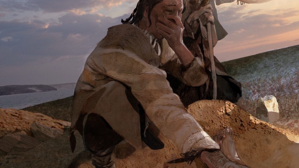
Ars Technica
TechnologyJun 18, 2026 · 1 min
Hunter-gatherers in Siberia died of a plague outbreak 5,500 years ago
Ars Technica
The Guardian
TechnologyJun 17, 2026 · 1 min
Ancient DNA provides evidence of earliest known plague outbreak
The Guardian

New Scientist
TechnologyJun 17, 2026 · 1 min
Oldest known plague outbreak killed hunter-gatherer children
New Scientist

CBC
NewsJun 17, 2026 · 1 min
Earliest outbreak of the plague killed hunter-gatherer kids 5,500 years ago
CBC